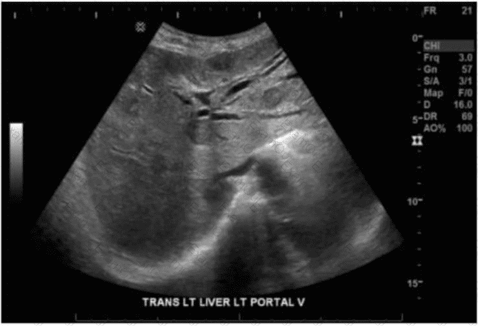
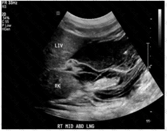
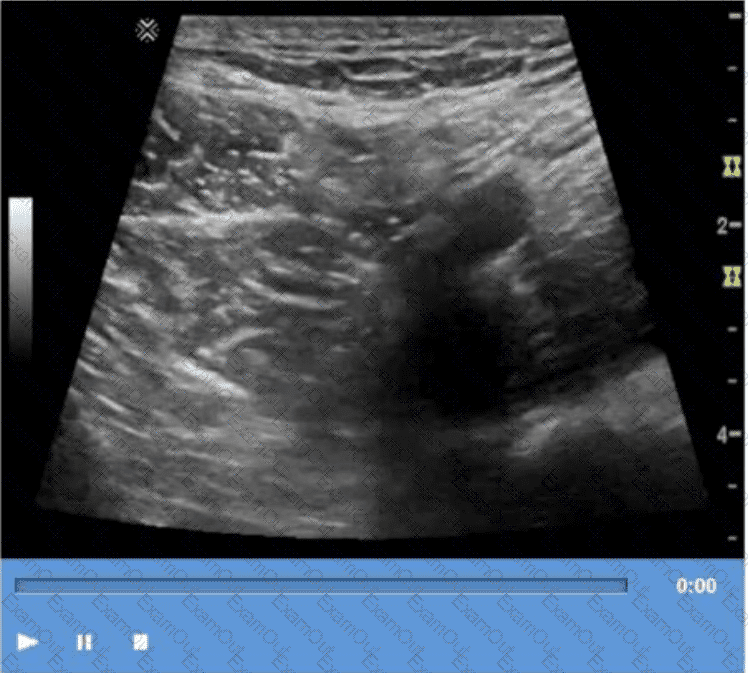
The technique shown in the video is compression. In ultrasound imaging—especially of soft tissue masses, the bowel, or venous structures—compression is used to evaluate the compressibility of structures. The image demonstrates a classic grayscale ultrasound view of a lesion or structure being compressed with the probe.
Compression sonography is particularly important in:
Evaluating venous patency (e.g., for deep vein thrombosis)
Differentiating cystic from solid structures
Evaluating bowel wall abnormalities or intussusception
Assessing lymph nodes and soft tissue masses (as shown here)
When a structure compresses easily under probe pressure, it suggests that the lesion is fluid-filled or soft. In contrast, incompressibility may indicate a solid mass or thrombus.
Differentiation from other options:
B. Valsalva: Involves forced expiration against a closed airway, used primarily to assess venous reflux or inguinal hernias—not what is demonstrated here.
C. Exhalation: A respiratory maneuver that passively alters thoracoabdominal pressure, not actively performed by the operator or causing focal structural change.
D. Deep inspiration: Used to improve visualization of the liver, diaphragm, or gallbladder—not to evaluate the compressibility of soft tissue.
[References:, Rumack CM, Wilson SR, Charboneau JW, Levine D. Diagnostic Ultrasound. 5th Edition. Elsevier, 2018. Chapter: Ultrasound Technique and Physics, pp. 35–39., AIUM Practice Parameter for the Performance of a Diagnostic Ultrasound Examination, 2020., , ]