Thin-layer chromatography has the advantage of being able to test for a large number of drugs at the same time.
Thin-layer chromatography is particularly useful as a tool in the identification of:
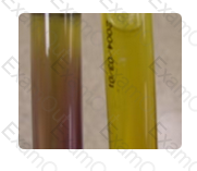
Conversion of only the slant to a pink color in a Christensen's urea agar slant is produced by bacterial species that have weak urease activity. The reaction in the slant to the right is often produced by Klebsiella species, as an example. Strong urease activity is indicated by conversion of the slant and the butt of the tube to a pink color, as seen in the tube to the left. The slant only reaction in the right tube may be seen early on if only the slant had been inoculated; however, with a strong urease producer, both the slant and the butt would turn. Therefore, the reaction is dependent on the strength of urease activity. If the media had outdated for a prolonged period, either there would be no reaction or the appearance of only a faint pink tinge, either in the slant, the butt or both, again depending on the strength of urease production by the unknown organism.
The urease reaction seen in the Christensen's urea agar slant on the far right indicates:

Sterile yellow stopper tubes contain thixotropic gel and a clot activator.
Question options:
Limited to blood and body fluids visibly contaminated with blood
Listeria monocytogenes is the correct answer. The motility agar is showing motility at the top of the tube, but not deeper; typical of this catalase-positive, gram positive bacillus. Streptococcus agalactiae would be catalase negative and a coccus. Erysipelothrix rhusiopathiae would be H2S-positive and catalase negative. Escherichia coli is a gram negative bacillus.
This Gram-positive bacillus grew as a diffusely beta-hemolytic colony from a newborn. It was catalase positive and had tumbling motility on a hanging drop preparation. This is how it appeared on triple sugar iron agar and motility medium. What is the most likely diagnosis?
Allergen-specific IgE, synthesized in response to allergens, becomes fixed to receptors on cellular membranes, especially those of basophils. If these receptor-bound IgE molecules are aggregated on re-exposure to specific allergen, both mast cells and basophils produce mediators that result in the allergic response. IgE-antigen interaction at the cell surface causes degranulation of cells and release of substances including: histamine, SRS-A, platelet activator, a kallikrein, and an eosinophil chemotactic factor. Basophils are the principal cells that bind IgE antibody while their number of receptor sites is proportional to serum IgE levels. Eosinophils are drawn to the site by the basophil chemotaxis mechanism, but are not the main cell which binds the IgE antibody.
Immunology
The mediator cells that bind MOST to IgE antibodies are:
Patients with antibody to the following antigen are immune to Hepatitis B:
Primary- Target glands (such as thymus, thyroid, parathyroid, etc.)
Secondary- Pituitary gland
Tertiary- Hypothalamus
Match the type of endocrine dysfunction with the appropriate organ:
1. Target gland
2. Pituitary gland
3. Hypothalamus
Rule-out is a process by which antibodies are identified as being unlikely in a given sample because of the absence of an expected antigen-antibody reaction. In other words, the absence of a reaction is noted with a cell that is positive for the corresponding antigen.
Although rule-out procedures may vary somewhat from institution to institution, the following general principles apply:
Non-reactive cells are selected for rule-out. To be classified as non-reactive, a cell must NOT have reacted at any phase of testing in a given panel or screen.
Using the logic that if the rule-out cell is positive for a given antigen, it should have reacted with the corresponding antibody, you can rule-out antibodies that correspond to antigen positive cells.
To increase the probability that rule-out will not mistakenly eliminate a weakly-reacting antibody that exhibits dosage*, use only cells that are homozygous for the corresponding antigen for those systems that generally show dosage. Generally these include: C, c, E, e, Fya, Fyb, Jka, Jkb, M, N, S, and s.
In this case, it is only possible to rule out on screening cell 2 since it demonstrates a negative reaction with the patient serum. Anti-C cannot be ruled out since the C antigen is heterozygous on screening cell 2 with c. Anti-Fya cannot be ruled out since this antigen is not present on screening cell 2. Anti-M and anti-Jka can be ruled out since the antigens are homozyous while demonstrating a negative reaction on screening cell 2.
Rule-out, while very useful, can lead to error. Ruling out an antibody should be combined with other supporting data to increase confidence in the solution; the more data collected, the higher the probability that the final solution is correct.
*Dosage means that there are two "doses" of the same antigen present on the red cells . Antibodies that exhibit dosage react more strongly with homozygous cells (e.g., Jka Jka) than with heterozygous cells (e.g., Jka Jkb) .
Based on the phenotype of the RBC screening cells, and patient results shown on the right, which of the following antibodies CANNOT be ruled out?
The activities conducted in a laboratory with a certificate of waiver include:
Any oxidase-positive organism can be excluded from the Enterobacteriaceae. The other characteristics are as a rule present in these organisms.
Which of the following is not true about members of the Enterobacteriaceae:
The Needlestick Safety and Prevention Act requires employers to:
Match the viruses below with their associated conditions.
1. Herpesvirus
2. Papovavirus
3. Rhinovirus
4. Rotavirus
Which of the following cardiac biomarkers rises within 30 minutes - 4 hours after chest pain, peaks in 2 - 12 hours, and is usually normal within 24 - 36 hours.
The approximate volume of CSF in an adult is 90-150 mL.
What is the approximate volume of spinal fluid in an adult?